
PaX-i3D Smart
Your first Partner for 3D Diagnosis
1 scan, 2 images (2D + 3D)
Innovative compressed Technology
Extended ARC shaped FOV
Product Highlights
Pax-i3D Smart
1 Scan, 2 Images
One scan with the Smart gives you not just a CT image but also an Auto Pano image.
This means patients who require both images do not need to undergo two X-ray scans.
Also, CT and Auto Pano images are displayed within the One Viewer feature.

Practical 3 in 1 System
CBCT ( with Auto Pano)
Panoramic
Cephalometric
Smart Innovation for Low Dose
1 Scan, 2 Images
Low-dose and High Images Quality
SMART Innovations for accurate diagnosis
- Anatomical FOV (12 x 9)
- ART-V
Anatomical FOV (12 x 9)
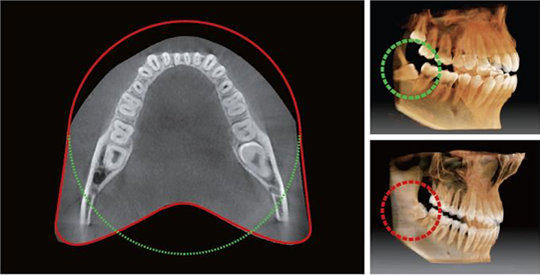
The innovative FOV of the PaX-i3D Smart provides an arch-shaped volume, which shows a wider view of dentition compared to other devices of the same FOV. Normally, a FOV 10x8.5 image shows tooth #8. However, when the tooth is lying on its side, there is a high possibility that the tooth will be cut out of the image. The "arch-shaped volume" eliminates this possibility and shows the hidden dentition are
ART-V (ARTIFACT REDUCTION TECHNOLOGY)by VatechART-V finds out the location of metals when radiation is exposed. It deletes the metal in projection data artificially and fills that part with similar values to the surrounding area. At the same time, it remembers the original location and status of metals and then it paints the area with a new value that has no streaks.
" Metal artifact hinders visualization and naturally reduces diagnostic confidence"

ART-V OFF

ART-V ON
PROFESSIONAL DIAGNOSTIC VALUE WITH PANORAMIC IMAGES
Smart Plus Provides the most precise and high-quality panoramic image. The clear and sharp panoramic image brings you better diagnostics. Enhanced details especially in the anterior and dental roots can be viewed.
These consistently high-quality images will become the new standard of panoramic imaging.

Magic PAN

Normal PAN
MAGIC PAN creates a more superb panorama image. It is acquired through the elimination of distorted and blurred images caused by improper patient positioning(Optional). Focused image is reorganized throughout the whole dental arch and the image quality can be increased. The image becomes clearer especially in the incisor and canine region, TMJ areas and root canal.
SCAN TYPE CEPHALOMETRIC
Scan type cephalometric offers two image sizes, LAT and FULL LAT, you can choose one of them based on the purposes of your diagnostic needs.
Lateral vs Full lateral
Provide specialized high quality images to suit Full lateral gives 30% larger images and the occipital orthodontics and maxillofacial surgeries' area of the patient for comprehensive diagnosis (optional).

Lateral
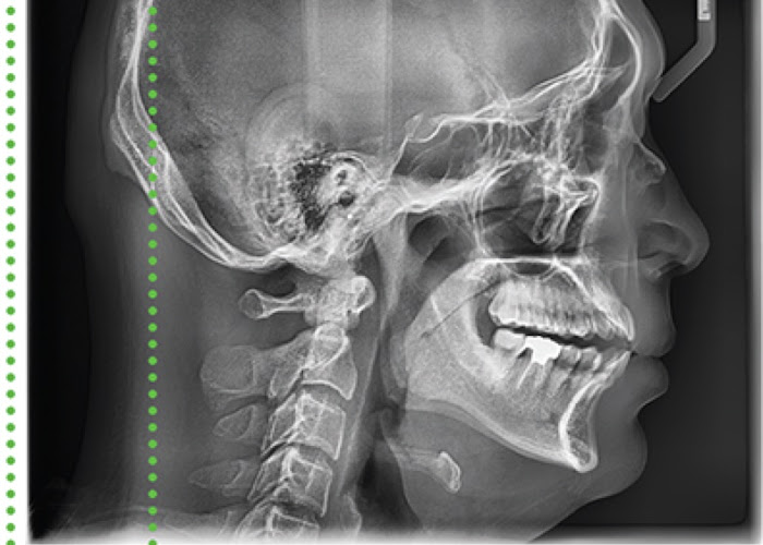
Full Lateral
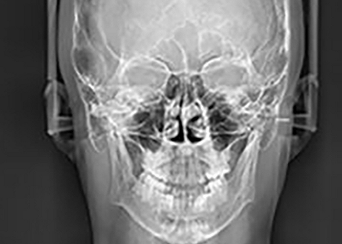
PA
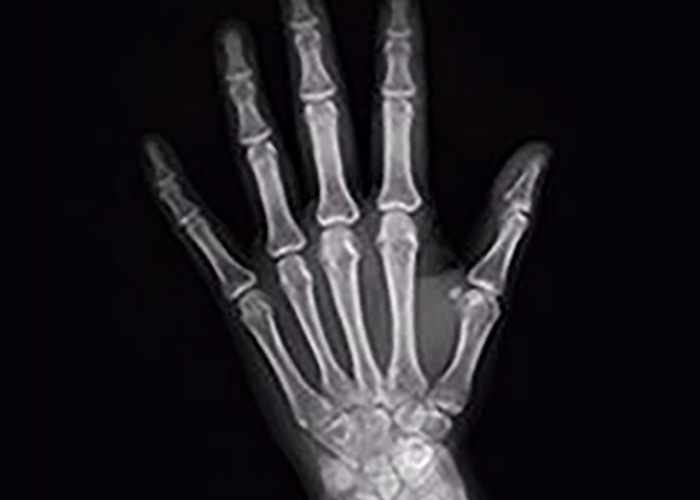
Carpus

SMV (Submentovertex)
Product Configuration & Specificatioins

Dimensions (Unit : mm)
